TM-Microscopy 4.98: User-friendliness and extended compatibility
TM-Microscopy 4.98: User-friendliness and extended compatibility
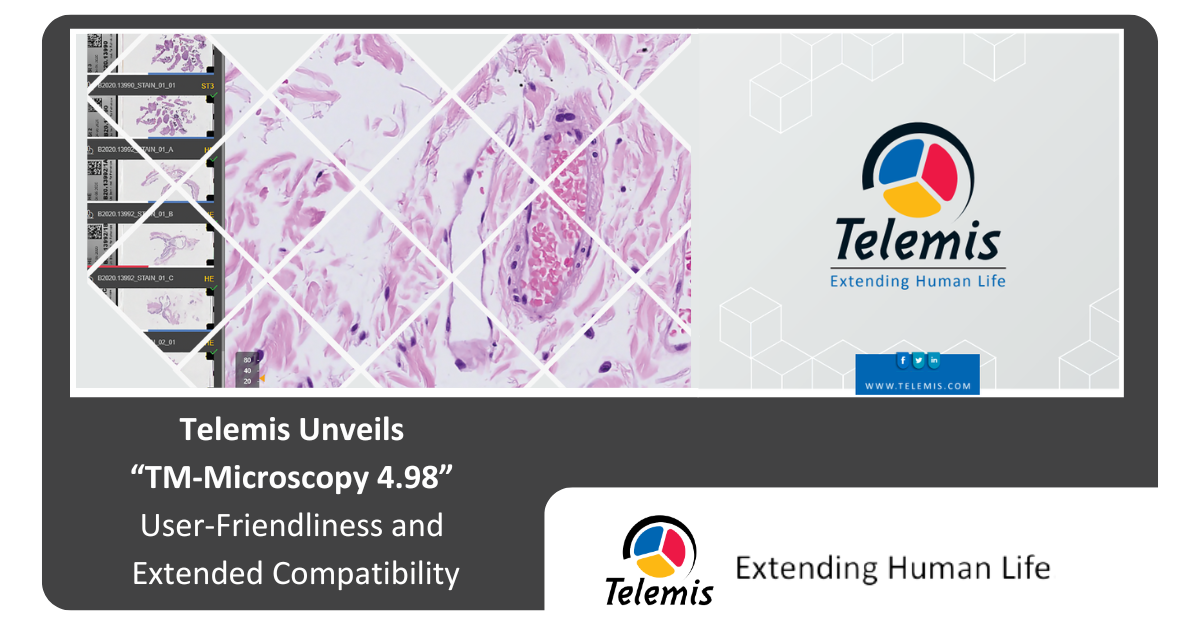
Louvain-la-Neuve, 27 May 2025. Telemis, the specialist in medical imaging solutions, is proud to announce TM-Microscopy 4.98. With this new version, TM-Microscopy takes a significant step forward and positions itself as an essential tool for digital pathology. Integration of the DICOM-WSI format, ultra-smooth navigation, extended annotations to macro images, and precise calibration of measurements: each development addresses a specific professional need. The goal is clear: to increase performance on-site and remotely.
DICOM-WSI: 100% compatible
The DICOM-WSI (Whole Slide Image) microscopic image format is gradually becoming an essential standard. To support this transition, TM-Microscopy significantly enhances its compatibility with this format. Integrations are now validated with Leica, Evident, and Excilone formats, ensuring optimal support for these standards. This standardisation considerably improves the compatibility and interoperability of microscopic slide images within medical data management systems.
A more user-friendly interface
In order to optimise the user experience and facilitate the daily work of professionals, the 4.98 version introduces several ergonomic improvements. The personalisation of keyboard shortcuts allows each user to adapt the tool to their preferences and working habits. Visual comfort is also at the user's choice through the selection between a dark or light interface. A new interface has been developed to facilitate communication between doctors and laboratory technicians, ensuring a smooth exchange of information and enhanced collaboration.
Calibration and annotations of macroscopy images
Annotations are now extended to macroscopic images. This feature offers increased flexibility to pathologists in their analysis and diagnostic work. They can now add comments, markings, measurements, and other indications directly on the sample images, thereby optimising sample tracking, the communication of observations, and case documentation.
Finally, by placing a reference ruler at the time of capture, the user can now calibrate the macroscopy images. This feature allows the direct measurement of tissue size in centimetres.
All these new annotation functions are available remotely, to enable faster support without compromising on the quality or accuracy of exchanges.
Conclusion
With version 4.98, TM-Microscopy continues to establish itself as the reference solution for digital pathology. By fully integrating medical imaging standards, streamlining daily use, and enhancing analysis and collaboration capabilities, this new version aligns with the current challenges faced by pathologists.